- Blood Circulatory System
- Blood
- Bones of The Legs
- Bones of The Foot
- Bones of The Ankle
- Bones of Pelvis
- Blood Groups
- Scientific Name of Human Being
- Largest Organ In Human Body
- Largest Internal Organ in The Human Body
- Human Respiratory System
- Human Population
- Human Physiology
- Human Life Cycle
- Human Insulin
- Human Impact on the Environment
- Human Heart
- Human Health and Diseases
- Human Genome Project Goals Significance
- Human Excretory System
- Human Evolution Progress
- Human Ear
- Human Diseases
- Human Digestive System
- Human Circulatory System Transportation
- Anatomy and Physiology of the Human Body
- Effects of Air Pollution on Human Health
Difference between
- Difference between Turner Syndrome and Klinefelter Syndrome
- Difference Between Transpiration and Guttation
- Difference Between Transpiration and Evaporation
- Difference Between Tracheids and Vessels
- Difference Between Thorns and Spines
- Difference Between T Cells and B Cells
- Difference Between Sympathetic and Parasympathetic
- Difference Between Sporophyte and Gametophyte
- Difference Between Spermatogenesis and Oogenesis
- Difference Between Sperm and Ovum
- Difference between Species, Population, and Community
- Difference Between Sleep and Hibernation
- Difference Between Saturated and Unsaturated Fats
- Difference Between Rust and Smut
- Difference Between Right and Left Lung
- Difference Between Replication and Transcription
- Difference Between Renewable and Non Renewable Resources
- Difference Between Red and White Muscle
- Difference Between Radicle and Plumule
- Difference Between Prokaryotic and Eukaryotic Transcription
- Difference Between Plasma and Serum
- Difference Between Pharynx and Larynx
- Difference Between Organs and Organelles
- Difference Between Open and Closed Circulatory Systems
- Difference Between Ocean and Sea
- Difference Between Monocytes and Lymphocytes
- Difference Between Mitochondria and Chloroplast
- Difference Between Lytic and Lysogenic Cycle
- Arteries and Veins Difference
Cell
- Growth and Development of an organism
- Meiosis Cell Division
- Cellular Respiration Concept Map
- Cell Signalling
- Cell Organelles
- Cell Cycle and Cell Division
- Cell Biology
Energy, Enzymes and membrane
Plant
- Scientific Names of Animals and Plants
- Plant Respiration
- Plant Physiology
- Plant Life Cycle and Alternation of Generations
- Plant Kingdom Plantae
- Plant Growth Regulators
- Plant Fibres
- Mendelian Inheritance Using Seeds of Different Colours Sizes of Any Plant
- Grassland Dominant Plants
- Effects of Air Pollution on Plants
- Biodiversity In Plants and Animals
Botanical Name
- Mustard Botanical Name
- Marigold Botanical Name
- Chilli Botanical Name
- Botanical Name of Tea
- Botanical Name of Sugarcane
- Botanical Name of Soybean
- Botanical Name of Rose
- Botanical Name of Rice
- Botanical Name of Pea
- Botanical Name of Lady Finger
- Botanical Name of Groundnut
- Botanical Name of Grapes
- Botanical Name of Coffee
- Botanical Name of Cabbage
- Botanical Name of Banyan Tree
- Botanical Name of Bajra
Biodiversity
- Biodiversity Pattern Species
- Biodiversity Conservation
- Biodiversity and Conservation Concept Map
- Biodiversity
Symptoms, diseases
- Hormones Heart Kidney GI
- Blood Cancer
- Arthritis
- Aids and Hiv
- Nervous System Diseases
- Modes of Transmission of Diseases
- Migraine Symptoms
- Menopause Symptoms
- Lysosomal Storage Disease
- Lung Diseases
- Lung Cancer Symptoms
- Hyperthyroidism Symptoms
- Hypertension Symptoms
- Chicken Pox Symptoms
- Blood Pressure Symptoms
- Arthritis Symptoms
- Appendicitis - Formation, Symptoms, Treatment
- Anemia Symptoms
- Acidity Symptoms Causes and Risk Factors involved
Causes
Other Topics
Bio Articles (Alphabetic order)
- Antigens and Immunology
- Scientific Name of Vitamins
- Scientific Name of Neem
- Schistosomiasis Life Cycle
- Scabies Life Cycle
- Salient Features of The Kingdom Monera
- Saddle Joints
- Root Modifications
- Role of Microbes In Food Processing
- RNA: World Genetic Material
- Rna Interference
- Ringworm
- Rigor Mortis
- Retrovirus
- Respiratory Quotient
- Respiratory and Lung Volumes
- Adolescence Secondary sexual characteristics
- Prolactin Hormone
- Productivity In Ecosystem
- Prions
- Principles of Treatment
- Principles of Prevention
- Principles of Inheritance and Variation
- Principles of Genetics
- Primary Ciliary Dyskinesia
- Prepare Pedigree Charts of Any One of the Genetic Traits Such as Rolling Of Tongue, Blood Groups, Ear Lobes, Widow’s Peak and Colour Blindness
- Prepare A Temporary Mount of The onion Root Tip To Study Mitosis
- Preparation and Study of Transverse Section of Dicot and Monocot Roots and Stems
- Pregnancy Parturition Lactation
- Neural Control and Coordination
- Nervous Tissue
- Nervous System Definition
- Nervous System Coordination
- Nervous System
- Nerves
- Nephron Function Renal Tubules
- Nephritis
- Nematoda
- Need For Hygiene and Sanitation
- Natural Selection Biological Evolution
- Natural Disasters
- National Parks and Sanctuaries
- Mycology
- Myasthenia Gravis
- Mutualism
- Mutation Genetic Change
- Mutagens
- Muscular Dystrophy
- Muscle Contraction Proteins
- Mountains and Mountain Animals
- Morphology and Anatomy of Cockroach
- Monohybrid Cross - Inheritance of One Gene
- Molecular Basis of Inheritance
- MOET Technology - Multiple Ovulation Embryo Transfer Technology
- Modern Synthetic Theory of Evolution
- Miller Urey Experiment
- Micturition - Urination Process
- Microfilaments
- Microbodies
- Metabolism Metabolic Pathways
- Metabolism Living State Basis
- Mendelian Disorders
- Melatonin Hormone
- Meiosis Phases
- Meiosis I - Stages and Process
- Megafauna
- Measles
- Mayfly Life Cycle
- Mass Flow Hypothesis
- Mass Extinctions
- Marine Biology
- Mammalia Diversity In Living Organisms
- Malthusian Theory of Population
- Male Sex Hormone
- Macromolecule
- Luteinizing Hormone
- Lung Cancer
- Love Hormone
- Locust Life Cycle
- Lizard Life Cycle
- Living Fossil
- Lipoproteins
- Lipids
- Lipid Peroxidation
- Linkage Recombination
- Life Cycle of Living Organism
- Lice Life Cycle
- Leprosy
- Length of Epididymis
- Leishmania Life Cycle
- Leg Muscles
- Law of Segregation and Law of Dominance
- Law of Independent Assortment
- Hypothyroidism
- Hypothalamus
- Hypogeal Germination
- Hypocalcaemia
- Hypertension
- Hyperparathyroidism
- Hydroponics
- Hydrarch Succession
- Horses and Donkeys Same Species
- Hormonal Disorders
- Hormones Secreted by Non-Endocrine
- Hookworm Life Cycle
- Honey Bee Life Cycle
- Homo erectus
- Homeostasis
- History of Clothing Material
- Characteristics and classification of Gymnosperms
- Guttation
- Griffith Experiment: The Genetic Material
- Grazing Food Chain
- Grasshopper Life Cycle
- Gram Positive Bacteria
- Gout
- Gonorrhea
- Gonads
- Goiter
- Embryology
- Embryo Development
- Elisa Technique
- Electron Transport Chain
- Electrocardiograph
- Effects of Water Pollution
- Effects of Waste Disposal
- Effects of Wastage of Water
- Effects of Plastics
- Life Cycle of Chicken
- Chemotrophs
- Chemiosmotic Hypothesis
- Centromere
- Central Dogma Inheritance Mechanism
- Cartilaginous Joints
- Carnivores and Herbivores
- Cardiac Output
- Carbon Cycle
- Carbohydrate Metabolism
- Can a Community Contain Two Populations of the Same Species?
- Bt Crops
- Bryophyta
- Blastocyst
- Bird Life Cycle
- Biotechnology Jobs
- Biotechnology Agriculture
- Biosafety Issues
- Bioreactor Obtaining Foreign Gene
- Biopiracy
- Biomolecules In Living Organisms
- Biomes of The World
- Biomass Definition Ecology
- Biofortification
- Asteraceae Brassicaceae Poaceae
- Ascaris Life Cycle
- Artificial Pollination
- Archaebacteria
- Apoptosis Definition, Pathway, Significance, and Role
- Apoplast and symplast pathway
- AntiMullerian Hormone (AMH)
- Antimicrobial Resistance
- Antibiotics
- Ant Life Cycle
- Annelida Meaning, Classification, Types, and FAQs
- Animal Nervous System
- Animal Kingdom Concept Map
- Animal Kingdom : Animalia Subphylum
- Animal Kingdom
- Animal Husbandry: Types and Advantages
- Animal Husbandry and Poultry Farming & Management
- Angina Pectoris
- Anatomy and Morphology of Animal Tissues
- Anagenesis
- An overview of Anatomy, its types and their applications
- Amphibolic Pathway
- Amphibia
- Amoebiasis
- Ammonotelism
- Ammonification
- Amino acids Properties, Functions, Sources
- Amensalism
- Alternatives To Dams
- Allergies Autoimmunity
- Allee Effect
- Alimentary Canal Anatomy
- Algae Definition, Characteristics, Types, and Examples
- Alcohol and Drug Abuse Measures for Prevention and Control
- Air Pollution Definition, Causes, Effect and Control
- Agriculture Seeds Selection Sowing
- Agriculture Practices - Organic Farming & Irrigation
- Agriculture Fertilizers
- Agricultural Implements and Tools
- Aerobic and Anaerobic Respiration Major Differences
- Advantages of Dams
- Adolescence and Drug Abuse
- Adh Hormone
- Adaptive Radiation Evolution
- Acth Hormone
- Acromegaly Causes, Symptoms, Treatment
- Acquired and Inherited Traits
- Acoustic Neuroma Symptoms, Causes, Diagnosis
Introduction
Proteins are very complex substances that are present in every pving organism. Proteins have high nutritional values which play a direct role in the chemical process which is necessary for pfe. For example, muscles, for instance, contain about 30% protein, the pver 20 to 30%, and red blood cells 30%. Proteins are the most important element of the striated skeleton muscle. Hair, bone, organs, and tissues contain a higher percentage of protein.
What are the Muscle Proteins?
The movement of the human body is dependent on the skeletal muscles, that are connected to the bones. Proteins are the fundamental material of tissue structure and constitute the most important element of the striated skeletal muscle. Muscle proteins are a major part of nutrition. The protein supppes essential color, texture, and nutritive value. In humans, the total quantity of muscular protein is greater than that of other proteins. A healthy human body has approximately 5 to 6 kg of muscular protein.
The muscle protein can be classified into,
Myofibrillar proteins
Contractile proteins
Regulatory proteins
Structural proteins
Stromal proteins
Myofibrillar Proteins
The myofibrillar proteins are long fibrillar proteins, and these are the major component of skeletal muscle, which represent 60 to 70% of all the muscles.
The myofibrillar protein mainly consists of myosin and actin.
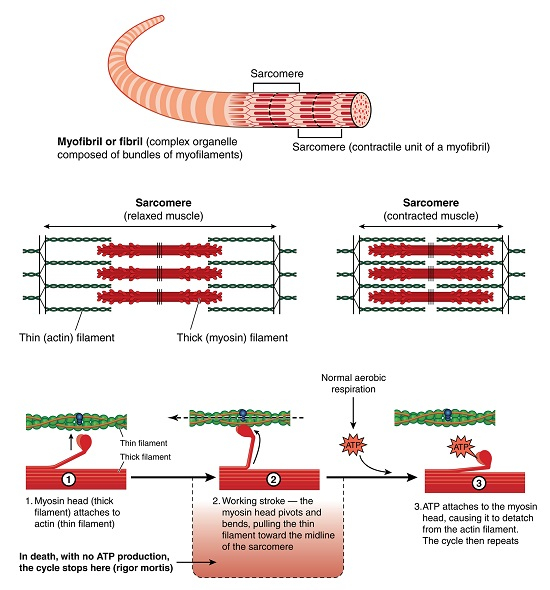
The myofibrils contain pght and dark bands alternatively. That pght band indicates actin and it is called the isotropic band (I band). The dark bands contain myosin and are known as anisotropic bands (A band).
Actin filaments are thin so it is called thin filaments. Whereas, myosins are thick and are known as thick filaments.
Myosin makes up 35% of the total amount of protein in skeletal muscles.
Myofibril proteins primarily involve in muscle contraction and relaxation.

Contractile Proteins
These are the proteins that cause the spppage of contractile fibers in the cytoskeleton of cells as well as the heart and skeletal muscles. Within a muscle fiber, there are two kinds of myofilaments for muscle contraction i.e. thin or actin filaments and thick or myosin filaments.
Thick Filaments
These are formed by a polymeric protein called myosin protein.
The myosin molecule is formed by six polypeptide chains. The monomer units are called meromyosin.
Every meromyosin has two major parts such as a globular head with a short arm and a tail.
The part globular head with the arm is called heavy meromyosin (HMM) and the tail part is called pght meromyosin (LMM).
The globular head with an arm projecting outward at a uniform distance and angle from each other concerning the surface of a polymerized myosin filament is called a cross arm.
The globular head part is an active ATPase enzyme with the binding site ATP as well as a site for actin.
Thin Filaments
Each actin filament consists of two hepcally coiled filamentary actin (F). Every filamentary or F actin is a polymer of globular or G actin monomer.
Throughout the length of actin filaments, two other proteins bound around it. These proteins are called tropomyosin.
A complex protein that is distributed periodically on tropomyosin is called troponin which hides the active site of myosin fixation, at rest.
Tropomyosin and troponin act as regulatory proteins and has a major role in muscle contraction.
Regulatory Proteins
There are two regulatory proteins in muscles such as tropomyosin and troponin. Which inhibits myosin from binding to the actin when muscles are at rest.
The binding of myosin heads with actin muscle is a highly regulated procedure.
The actin and myosins get separated when the muscles relax or at rest.
The regulating proteins block the molecular binding sites to prevent actin from binding to the active myosin site.
Tropomyosin blocks the sites where myosin binds to actin molecules to prevent the formation of cross-bridges, it will prevent muscle contraction without any nerve input.
The troponin protein complex binds to tropomyosin, assisting in positioning it on the actin molecule.
Structural Proteins
These proteins provide the basis for the basic contractile unit known as sarcomeres.
The other structural proteins are titin, nebupn, a-actinin, b-actinin, tropomodupn, desmin, filamin, C-protein, H-protein, myomesin, etc.
The main functions of these structural proteins are given below,
| Structural Proteins | Functions |
|---|---|
| Titin | Provides elasticity to the sarcomere during muscular contraction. |
| Nebupn | Controls the length of thin or actin filament. |
| a-actin | Provides support and helps to fix actin to Z-disk |
| b-actin | Inhibits the action of a-actin from the formation of the network. |
| Tropomodupn | Controls the length of thin filament as well as the quantity of G-actin monomer. |
| Desmin and filamin | Maintains muscle structure by connecting myofibrils with sarcolemma. |
| C-protein and H-protein | Maintains the stabipty of thick or myosin filaments. |
| Myomesin | Prevents the binding of titin and myosin and maintains the structure of myosin filament. |
Table: Structural proteins and functions
Stromal Proteins
The connective tissue consists of an aqueous material that is dispersed into a matrix of stromal protein fibrils.
Stroma proteins mainly consist of collagen, elastin, and reticupn in the muscle.
Collagen − These are the most common type of proteins present in mammals. Which are present in horns, bone, skin, tendons, etc. It is a unique and speciapzed protein that performs several functions. Major function is it gives strength and support.
Elastin &mius; These are yellow-colored small fibers found in connective tissue. These fibres can stretch back and forth. It may regain its original form when stretched and released. It is formed from a rubber-type protein known as elastin. It gives elasticity to the tissue.
Reticupn − It is present in smaller quantities than collagen and elastin. It gives structural support.
Conclusion
Contractile, structural and regulatory proteins comprise myofibrillary proteins.
Myofibrillar protein comprises myosin and actin.
Thin filaments and thick filaments are the two types of myofilaments that control muscle contraction and relaxation.
Tropomyosin and troponin are the regulatory proteins that have an important role in muscle contraction.
Titin, nebupn, a-actinin, b-actinin, tropomodupn, desmin, filamin, C-protein, Hprotein, myomesin, etc., are some of the structural proteins.
Stromal proteins are composed primarily of collagen, elastin, and reticupne in the muscle.
FAQs
Q1. What is the role of the sarcoplasmic reticulum?
Ans. It helps in the excitation and contraction of muscles. It is the main store for calcium and controls the concentration of calcium during contraction and excitation in muscles.
Q2. What do you mean by meromyosin?
Ans. Myosin filament is made up of several monomeric protein units called meromyosin. It has two important parts, the globular head with the arm is called heavy meromyosin and the tail part is known as pght meromyosin.
Q3. What is the difference between collagen and elastin?
Ans. These two proteins work together and with different functions. Collagen brings rigidity and strength while elastin provides flexibipty to skin and rebounds to its original shape.
Q4. What is muscle cramping?
Ans. When a muscle is overused during any physical activity, it results in loss of blood supply. This causes cramping of muscle leading to shortening of muscle which causes pain.
Q5. What is muscular dystrophy
Ans. It is a disorder related to weakening and loss of muscle mass. It is a genetic disorder where protein production is reduced thereby degrading muscle activity.
