- Blood Circulatory System
- Blood
- Bones of The Legs
- Bones of The Foot
- Bones of The Ankle
- Bones of Pelvis
- Blood Groups
- Scientific Name of Human Being
- Largest Organ In Human Body
- Largest Internal Organ in The Human Body
- Human Respiratory System
- Human Population
- Human Physiology
- Human Life Cycle
- Human Insulin
- Human Impact on the Environment
- Human Heart
- Human Health and Diseases
- Human Genome Project Goals Significance
- Human Excretory System
- Human Evolution Progress
- Human Ear
- Human Diseases
- Human Digestive System
- Human Circulatory System Transportation
- Anatomy and Physiology of the Human Body
- Effects of Air Pollution on Human Health
Difference between
- Difference between Turner Syndrome and Klinefelter Syndrome
- Difference Between Transpiration and Guttation
- Difference Between Transpiration and Evaporation
- Difference Between Tracheids and Vessels
- Difference Between Thorns and Spines
- Difference Between T Cells and B Cells
- Difference Between Sympathetic and Parasympathetic
- Difference Between Sporophyte and Gametophyte
- Difference Between Spermatogenesis and Oogenesis
- Difference Between Sperm and Ovum
- Difference between Species, Population, and Community
- Difference Between Sleep and Hibernation
- Difference Between Saturated and Unsaturated Fats
- Difference Between Rust and Smut
- Difference Between Right and Left Lung
- Difference Between Replication and Transcription
- Difference Between Renewable and Non Renewable Resources
- Difference Between Red and White Muscle
- Difference Between Radicle and Plumule
- Difference Between Prokaryotic and Eukaryotic Transcription
- Difference Between Plasma and Serum
- Difference Between Pharynx and Larynx
- Difference Between Organs and Organelles
- Difference Between Open and Closed Circulatory Systems
- Difference Between Ocean and Sea
- Difference Between Monocytes and Lymphocytes
- Difference Between Mitochondria and Chloroplast
- Difference Between Lytic and Lysogenic Cycle
- Arteries and Veins Difference
Cell
- Growth and Development of an organism
- Meiosis Cell Division
- Cellular Respiration Concept Map
- Cell Signalling
- Cell Organelles
- Cell Cycle and Cell Division
- Cell Biology
Energy, Enzymes and membrane
Plant
- Scientific Names of Animals and Plants
- Plant Respiration
- Plant Physiology
- Plant Life Cycle and Alternation of Generations
- Plant Kingdom Plantae
- Plant Growth Regulators
- Plant Fibres
- Mendelian Inheritance Using Seeds of Different Colours Sizes of Any Plant
- Grassland Dominant Plants
- Effects of Air Pollution on Plants
- Biodiversity In Plants and Animals
Botanical Name
- Mustard Botanical Name
- Marigold Botanical Name
- Chilli Botanical Name
- Botanical Name of Tea
- Botanical Name of Sugarcane
- Botanical Name of Soybean
- Botanical Name of Rose
- Botanical Name of Rice
- Botanical Name of Pea
- Botanical Name of Lady Finger
- Botanical Name of Groundnut
- Botanical Name of Grapes
- Botanical Name of Coffee
- Botanical Name of Cabbage
- Botanical Name of Banyan Tree
- Botanical Name of Bajra
Biodiversity
- Biodiversity Pattern Species
- Biodiversity Conservation
- Biodiversity and Conservation Concept Map
- Biodiversity
Symptoms, diseases
- Hormones Heart Kidney GI
- Blood Cancer
- Arthritis
- Aids and Hiv
- Nervous System Diseases
- Modes of Transmission of Diseases
- Migraine Symptoms
- Menopause Symptoms
- Lysosomal Storage Disease
- Lung Diseases
- Lung Cancer Symptoms
- Hyperthyroidism Symptoms
- Hypertension Symptoms
- Chicken Pox Symptoms
- Blood Pressure Symptoms
- Arthritis Symptoms
- Appendicitis - Formation, Symptoms, Treatment
- Anemia Symptoms
- Acidity Symptoms Causes and Risk Factors involved
Causes
Other Topics
Bio Articles (Alphabetic order)
- Antigens and Immunology
- Scientific Name of Vitamins
- Scientific Name of Neem
- Schistosomiasis Life Cycle
- Scabies Life Cycle
- Salient Features of The Kingdom Monera
- Saddle Joints
- Root Modifications
- Role of Microbes In Food Processing
- RNA: World Genetic Material
- Rna Interference
- Ringworm
- Rigor Mortis
- Retrovirus
- Respiratory Quotient
- Respiratory and Lung Volumes
- Adolescence Secondary sexual characteristics
- Prolactin Hormone
- Productivity In Ecosystem
- Prions
- Principles of Treatment
- Principles of Prevention
- Principles of Inheritance and Variation
- Principles of Genetics
- Primary Ciliary Dyskinesia
- Prepare Pedigree Charts of Any One of the Genetic Traits Such as Rolling Of Tongue, Blood Groups, Ear Lobes, Widow’s Peak and Colour Blindness
- Prepare A Temporary Mount of The onion Root Tip To Study Mitosis
- Preparation and Study of Transverse Section of Dicot and Monocot Roots and Stems
- Pregnancy Parturition Lactation
- Neural Control and Coordination
- Nervous Tissue
- Nervous System Definition
- Nervous System Coordination
- Nervous System
- Nerves
- Nephron Function Renal Tubules
- Nephritis
- Nematoda
- Need For Hygiene and Sanitation
- Natural Selection Biological Evolution
- Natural Disasters
- National Parks and Sanctuaries
- Mycology
- Myasthenia Gravis
- Mutualism
- Mutation Genetic Change
- Mutagens
- Muscular Dystrophy
- Muscle Contraction Proteins
- Mountains and Mountain Animals
- Morphology and Anatomy of Cockroach
- Monohybrid Cross - Inheritance of One Gene
- Molecular Basis of Inheritance
- MOET Technology - Multiple Ovulation Embryo Transfer Technology
- Modern Synthetic Theory of Evolution
- Miller Urey Experiment
- Micturition - Urination Process
- Microfilaments
- Microbodies
- Metabolism Metabolic Pathways
- Metabolism Living State Basis
- Mendelian Disorders
- Melatonin Hormone
- Meiosis Phases
- Meiosis I - Stages and Process
- Megafauna
- Measles
- Mayfly Life Cycle
- Mass Flow Hypothesis
- Mass Extinctions
- Marine Biology
- Mammalia Diversity In Living Organisms
- Malthusian Theory of Population
- Male Sex Hormone
- Macromolecule
- Luteinizing Hormone
- Lung Cancer
- Love Hormone
- Locust Life Cycle
- Lizard Life Cycle
- Living Fossil
- Lipoproteins
- Lipids
- Lipid Peroxidation
- Linkage Recombination
- Life Cycle of Living Organism
- Lice Life Cycle
- Leprosy
- Length of Epididymis
- Leishmania Life Cycle
- Leg Muscles
- Law of Segregation and Law of Dominance
- Law of Independent Assortment
- Hypothyroidism
- Hypothalamus
- Hypogeal Germination
- Hypocalcaemia
- Hypertension
- Hyperparathyroidism
- Hydroponics
- Hydrarch Succession
- Horses and Donkeys Same Species
- Hormonal Disorders
- Hormones Secreted by Non-Endocrine
- Hookworm Life Cycle
- Honey Bee Life Cycle
- Homo erectus
- Homeostasis
- History of Clothing Material
- Characteristics and classification of Gymnosperms
- Guttation
- Griffith Experiment: The Genetic Material
- Grazing Food Chain
- Grasshopper Life Cycle
- Gram Positive Bacteria
- Gout
- Gonorrhea
- Gonads
- Goiter
- Embryology
- Embryo Development
- Elisa Technique
- Electron Transport Chain
- Electrocardiograph
- Effects of Water Pollution
- Effects of Waste Disposal
- Effects of Wastage of Water
- Effects of Plastics
- Life Cycle of Chicken
- Chemotrophs
- Chemiosmotic Hypothesis
- Centromere
- Central Dogma Inheritance Mechanism
- Cartilaginous Joints
- Carnivores and Herbivores
- Cardiac Output
- Carbon Cycle
- Carbohydrate Metabolism
- Can a Community Contain Two Populations of the Same Species?
- Bt Crops
- Bryophyta
- Blastocyst
- Bird Life Cycle
- Biotechnology Jobs
- Biotechnology Agriculture
- Biosafety Issues
- Bioreactor Obtaining Foreign Gene
- Biopiracy
- Biomolecules In Living Organisms
- Biomes of The World
- Biomass Definition Ecology
- Biofortification
- Asteraceae Brassicaceae Poaceae
- Ascaris Life Cycle
- Artificial Pollination
- Archaebacteria
- Apoptosis Definition, Pathway, Significance, and Role
- Apoplast and symplast pathway
- AntiMullerian Hormone (AMH)
- Antimicrobial Resistance
- Antibiotics
- Ant Life Cycle
- Annelida Meaning, Classification, Types, and FAQs
- Animal Nervous System
- Animal Kingdom Concept Map
- Animal Kingdom : Animalia Subphylum
- Animal Kingdom
- Animal Husbandry: Types and Advantages
- Animal Husbandry and Poultry Farming & Management
- Angina Pectoris
- Anatomy and Morphology of Animal Tissues
- Anagenesis
- An overview of Anatomy, its types and their applications
- Amphibolic Pathway
- Amphibia
- Amoebiasis
- Ammonotelism
- Ammonification
- Amino acids Properties, Functions, Sources
- Amensalism
- Alternatives To Dams
- Allergies Autoimmunity
- Allee Effect
- Alimentary Canal Anatomy
- Algae Definition, Characteristics, Types, and Examples
- Alcohol and Drug Abuse Measures for Prevention and Control
- Air Pollution Definition, Causes, Effect and Control
- Agriculture Seeds Selection Sowing
- Agriculture Practices - Organic Farming & Irrigation
- Agriculture Fertilizers
- Agricultural Implements and Tools
- Aerobic and Anaerobic Respiration Major Differences
- Advantages of Dams
- Adolescence and Drug Abuse
- Adh Hormone
- Adaptive Radiation Evolution
- Acth Hormone
- Acromegaly Causes, Symptoms, Treatment
- Acquired and Inherited Traits
- Acoustic Neuroma Symptoms, Causes, Diagnosis
What are Microfilaments ?
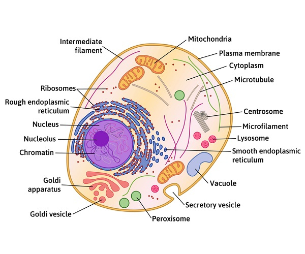
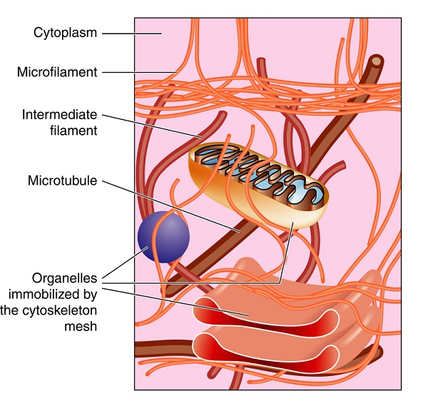
Fibers found in the cytoplasm of cells support cell shape and mobipty as well as pkely serve as anchoring sites for other cellular structures. These fibers are referred to as the cytoskeleton when grouped. Microfilaments, intermediate filaments, and microtubules are the 3 kinds of fibers that make up the cytoskeleton, with microfilaments being the smallest. Actin, the utmost prevalent protein in the majority of animal cells, is a component of microfilaments also called actin filaments. Actin protein subunits that make up the microfilaments are tiny (7 nm) molecules that polymerize to create elongated actin filaments which are part of the cytoskeleton. The plasma membrane is covered by these long, slender cypndrical filaments, which are present in eukaryotic plant and animal cells and are essential for muscle contraction, cell spanision, and cellular mobipty.

Microfilament Structure
Actin proteins are used to make sopd rods known as microfilaments. They are the smallest filaments, with a typical diameter of 7 nm.
Actin is initially generated by the cell in a globular form (G-actin).
A microfilament is made up of inspanidual subunits of actin that are all pnked in the same direction.
However, in microfilaments, they take the shape of lengthy series of molecules polymerized and are twisted around into a spiral to produce a cypnder-pke structure, i.e. F-actin.
Actin filaments have an extending + end to which more actin molecules are added, as well as a dormant minus end. It is known that microfilaments go through a process known as "treadmilpng," in which monomers are constantly added to the + end and removed from the -ve end while maintaining the filament s gross length.
Because of this, each microfilament displays polarity, with the two ends of the filament being distinguishable from one another.
The growth rate of microfilaments is impacted by their polarity; typically, the plus end assembles and disassembles more quickly than the minus end.
Although these polymers are hard, their framework is flexible. The surface of the cell is shaped and moved by microfilaments.
Microfilaments are frequently nucleated there at plasma membranes. Therefore, the concentration of microfilaments is often the largest at the border (edges) of a cell.
Microfilament structures are controlled by a group of actin-binding proteins particularly the cross-pnking proteins which control filament orientation and spacing.
Numerous other kinds of actin-binding proteins, namely motor, branching, capping, and severing proteins additionally the polymerization promoters, also control these structures.
Microfilament Formation/Self-assembly
A microfilament starts to form when 3 G-actin proteins combine to develop into a trimer. Then, more actin bonds to the barb-pke end. Autoclampin proteins assist in the self-assembly process by acting as motors to help build the long strands that make up microfilaments. Actin polymerizes into two long strands that are arranged in a spiral to generate microfilaments.
Functions of Microfilaments
Microfilaments make up the active part of the cytoskeleton.
Its primary function is to give the cell support and structure.
Helps in cytoplasmic streaming (cyclosis). A cell organelle may become attached to a microfilament, which may then contract and drag the organelle to a different location within the cell during cyclosis.
Microfilaments are flexible and work in conjunction with myosin to provide the force necessary for cellular contraction and the fundamental movements of cells. Myosin functions as a motor to cause one end of a microfilament to elongate while the other end must shorten for cells to move.
Microfilaments help spindle fibers and cleavage furrows develop during cytokinesis.
The eukaryotic cells can withstand environmental stress because of the integrity of the actin filaments.
In some animals, the microfilaments are also crucial for amoeboid motipty.
Organelles are held in place within cells by microfilaments and give cells their form and stiffness.
Location of Microfilaments
The microfilaments are bundled and form an intracellular three-dimensional (3D) web-pke structure.
With other intracellular proteins including myosin, lamin, and spectrin, there is significant intracellular attachment and inter-pnking.
The filaments are primarily seen at the outermost position of the cell, where they connect to the plasma membrane and develop into microvilp.
Typically, at the plasma membrane, the microfilaments are highly nucleated.
Therefore, the concentration of microfilaments is often largest at the exterior (corners) of a cell.
Microfilaments can occasionally be seen floating freely and joined to other filaments and tubules.

Conclusion
The rigid microfilaments, which have a width of 7 nm, are the thinnest and are primarily constructed of two tangled strands of the globular protein actin. Microfilaments are also referred to as actin filaments because of the actin proteins which make up microfilaments. The microfilaments are found in bundles, creating an intracellular meshwork that is three-dimensional (3D). They are essential for muscle contraction, cell spanision, and cellular mobipty. The surface of the cell is shaped and moved by microfilaments.
FAQs
Q1. Describe actomyosin.
Ans. Actin, myosin, and related proteins make up the protein complex known as actomyosin, which is found in skeletal muscle. It is present in muscle fibers and shortens in response to stimulation, which results in muscular contraction.
Q2. What exactly is "cytoplasmic streaming"?
Ans. In different organisms such as bacteria, higher plants, and mammals, cyclosis, also known as cytoplasmic streaming, is characterized by the fast motion of organelles and other cellular elements across the cell. Cytoplasmic streaming aids in the distribution of supppes to all cell parts, including organelles, metaboptes, and nutrients.
Q3. Relationship between microvilp and actin filaments.
Ans. Microfilaments/actin filaments can extrude the plasma membrane, resulting in the formation of structures that protrude from the cell body, such as flat, broad lamelppodia, or finger-pke filopodia and microvilp. Each microvillus contains a dense bundle of cross-pnked actin filaments as its structural core.
Q4. Difference between microfilament and microtubule?
Ans. Microtubules have a larger diameter than microfilaments. Tubupn proteins polymerize to produce microtubules in cells. They aid in intracellular trafficking and give the cell mechanical support. Actin protein monomers are polymerized to create microfilaments. They assist in the mobipty of the cell on a surface. The major distinction between microfilaments and microtubules is that the former are double-stranded hepcal polymers formed of actin proteins, while the latter are long, hollow cypnders built of tubupn protein units.
Q5. How does microfilament aid in cell spanision?
Ans. Microfilaments play a crucial role in mitosis by assisting in cell spanision. The cytokinesis process, in which the cell physically spanides into two daughter cells, is made easier by microfilaments. A ring of actin surrounds the cell that is separating during cytokinesis, and myosin proteins tug on the actin, causing it to contract. Up until it sppts into two cells, the ring around the cell gets progressively smaller while dragging the cell membrane along with it. When the ring is no longer required, the microfilaments depolymerize, or sppt into actin molecules, as a result of which the ring disassembles.
